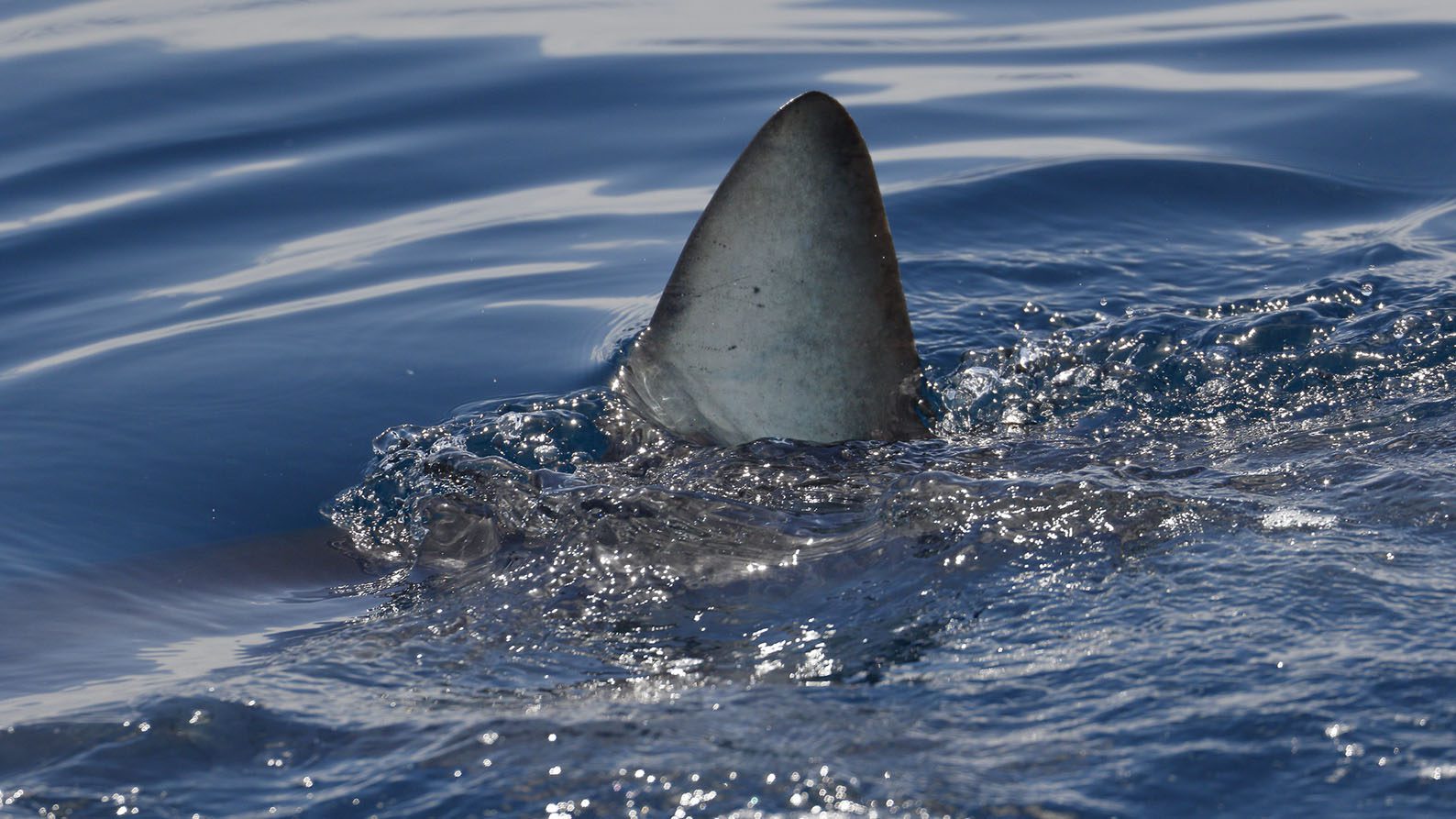
Curtin University researchers have revealed how the pelvic fins of fish such as sharks and chimaeras have evolved from their sudden appearance in the fossil record over 410 million years ago.
The team used CT scanning and 3D modelling to study the growth of pelvic fins in fish embryos to help us understand how the skeleton of these fins changed over evolutionary history.
Lead author and PhD candidate Jacob Pears from Curtin’s School of Molecular and Life Sciences said the research showed what the development of modern animals can tell us about their evolution.
“Our work focussed on cartilaginous fish and in particular looked at the pelvic fins of elephant sharks. The fine detail from our imaging revealed the basipterygium (pelvic fin bar), which like the femur and tibia in humans, were formed by the fusion of fin radials during early embryonic development,” Mr Pears said.
“In primitive sharks and cartilaginous fish, pelvic fin radials attach primarily to the pelvis while in more modern fish the radials are almost always found on the pelvic fin bar.
“Our findings suggests that the mechanism responsible for these alterations in the pelvic fin skeleton over millions of years is changes in where and how much the fin radials fuse together during early development.”
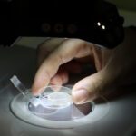
Shark embryos as small as 65mm were nano-CT scanned in collaboration with researchers at McGill University in Canada, with each part of the pelvic skeleton modelled in 3D at the Curtin HIVE (Hub for Immersive Visualisation and eResearch).
Visualisation technology specialist at the Curtin HIVE Carley Tillett said the CT scans were used to visualise and model the earliest stages of skeletal development of the elephant shark embryos.
“Our work illustrates how modern imaging technologies can provide insight into the development of modern animals and inform our understanding of the evolution of their anatomy,” Ms Tillett said.
The research was funded by an Australian Government Research Training Program scholarship, Curtin Research Fellowship, the Canadian Foundation of Innovation and the Australian Research Council (ARC) grant.
Published in the Journal of Developmental Biology, the research is titled ‘The Development of the Chimaeroid pelvic skeleton and the Evolution of Chondrichthyan Pelvic Fins’ and is available online here.